Instructions: Study all the visual information in this online lab, plus the accompanying explanations. Be prepared to answer a few "check-in" questions to show that you were paying attention. |
Muscle tissue | Smooth muscle tissue. |
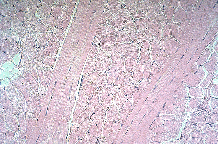
Skeletal (striated) muscle tissue. Some fibers are cut longitudinally, others transversely. | Muscle tissue striations. |
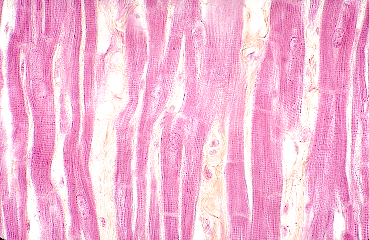
Cardiac muscle tissue: In this slide and the one to the right, notice the striations and the cell boundaries (intercalated disks) running perpendicular to the fiber direction; also notice the centrally located nuclei. | Cardiac muscle tissue |
Nerve cells and nervous tissue |
Cerebral hemispheres (cross-section) |
Spinal cord: gray matter (left) and white matter (right) |
Motor neuron cell bodies in ventral horn of spinal column |
Cell body of motor neuron |
Neuron cell body, with neurofibrils |
Peripheral nerves in cross-section |
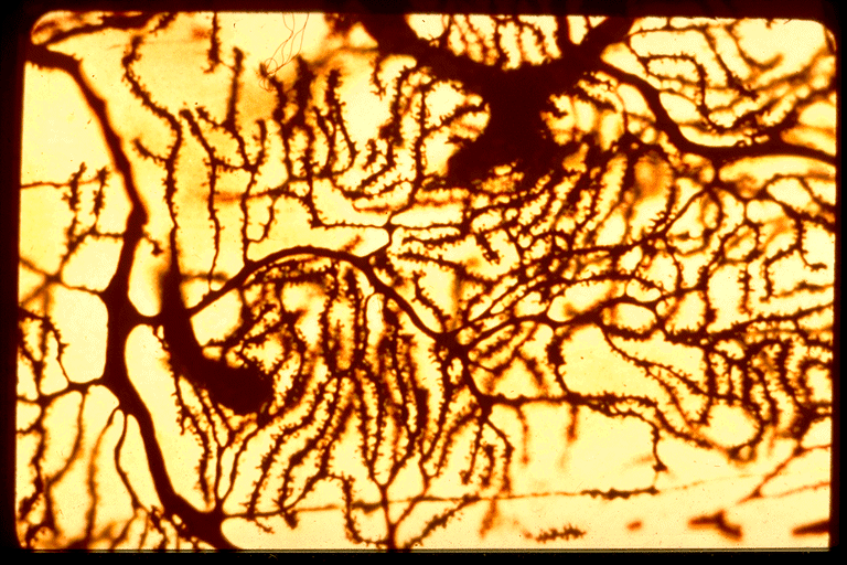
Dendrites |
Astrocytes |
Fibrous astrocyte |
Oligodendroglia |
Basket cell (large, right of center) and granule cells (smaller, with fewer branches) |
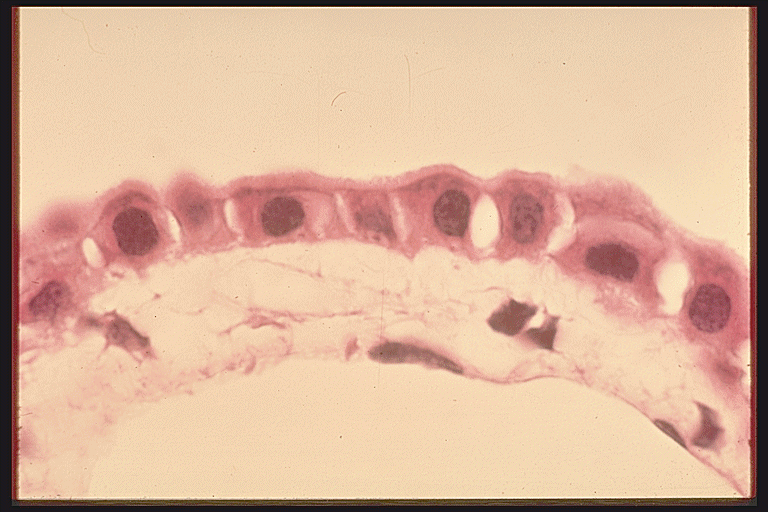
Simple squamous epithelium (on surface) |
Simple cuboidal epithelium |
Simple columnar epithelium |
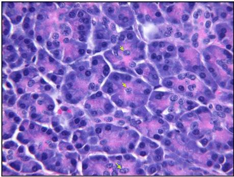
Glandular epithelium: pancreas (exocrine, acinar) |
Columnar epithelium (pseudostratified, ciliated) |
Transitional epithelium (collapsed) |
Transitional epithelium (stretched) |
Stratified squamous epithelium from skin (keratinized at the surface) |
Stratified columnar epithelium |
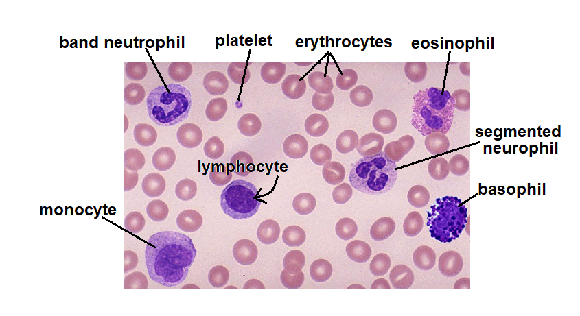
Blood, showing various cell types | |
Mesenchyme tissue |
Loose (areolar) connective tissue |
Reticular connective tissue |
Collagen fibers (wiggly bundles) and fat cells (circular, empty-looking) |
Dense, irregular connective tissue (with collagen fibers going in many directions) |
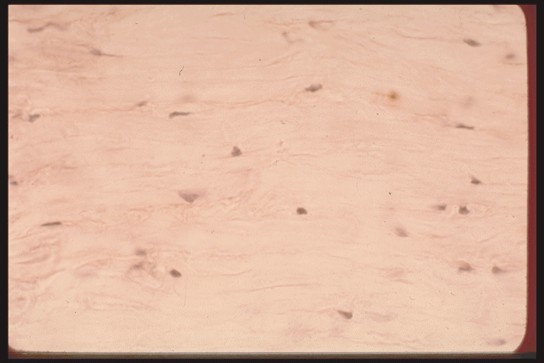
Dense, regular connective tissue (with collagen fibers all aligned) |